When it comes to patient care, our first responsibility is to gather as much information about you and your medical history so we can provide the best care possible. Check out the article below, written by medical assistant at Prairie Spine, Rebecca Thompson, to review the diagnostics available that investigate causes of pain and discomfort.
An important part of seeing a spine surgeon is imaging. Imaging comes in several different forms and each show things a little differently.
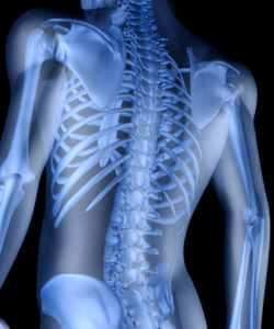 X-rays are the most common imaging study done and show the “bony anatomy” the best. X-rays do vary depending on whether they are done standing or laying down. The providers here at Prairie spine require standing images to see what gravity does to your spine.
MRI’s (Magnetic Resonance Imaging) show more of the soft tissue anatomy. Bones are shown in MRI as well, but this is the best study to see things like nerve impingement, disc bulges and the soft tissue changes often associated with spine pain.
CT’s (Computed Tomography) can show both bone and soft tissue, but do not show soft tissue such as nerves and disc bulges with as much clarity and detail as an MRI. However, for those unable to have MRI’s a CT Myelogram is often the only way to see disc bulges and nerve impingement.
So, don’t be surprised if the doctor requires more or a different kind of imaging than you have previously had. The most important thing for a surgeon is making sure he has all of the needed information to treat each individual as just that, an individual. Having the full picture allows the doctor to do the least invasive and most appropriate surgery possible!
Here is a video that shows the anatomy of the spine.
Our experts are a phone call away at 309.691.7774.